

By Dr. Philemon Huang, MBBS, MMed (Ophth), FAMS | Senior Consultant Ophthalmologist
Medically Reviewed by Dr. Leslie Ang Pek Seng, MBBS, MMed (Ophth)
Last Updated: June 2026
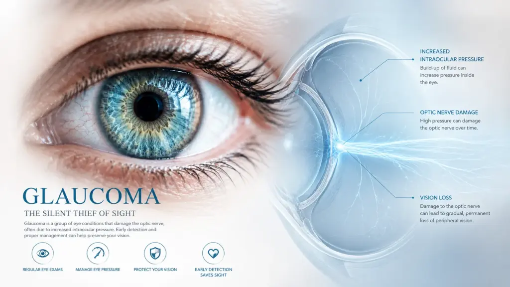
The Quick Answer: Glaucoma is a progressive, irreversible disease that damages the optic nerve, often due to high eye pressure. Because it destroys peripheral vision first, up to 40% of vision can be lost before a patient notices symptoms. Management includes medicated drops, Selective Laser Trabeculoplasty (SLT) laser therapy, Minimally Invasive Glaucoma Surgery (MIGS) and glaucoma filtration surgeries/drainage devices like Trabeculectomy and Tubes. MediSave can be utilized for laser and surgical interventions in Singapore.
Why Standard Screenings Miss Early Glaucoma
Glaucoma is the leading cause of irreversible blindness worldwide. In our clinical practice at Mount Elizabeth Medical Centre Orchard, the most tragic cases we encounter are not those that are complex to treat, but those that present too late.
Many patients assume that if they can read the 6/6 (or 20/20) line on an eye chart at the optical shop, their eyes are healthy. This is an erroneous assumption. Glaucoma is a disease of the optic nerve, not the cornea or the lens. It quietly erodes your peripheral (side) vision over years. By the time it affects your central vision—the part used to read messages on your phone—the damage is permanent.
Relying solely on a basic “air-puff” eye pressure test is insufficient. To comprehensively rule out glaucoma, we must perform Visual Field Testing (perimetry) and an Optical Coherence Tomography (OCT) scan of the optic nerve head. This allows us to pick up early visual field losses and measure the thickness of the Retinal Nerve Fiber Layer (RNFL) and Ganglion cell inner plexiform layer (GCIPL) down to the micron, detecting structural loss before you notice visual symptoms.

Understanding Intraocular Pressure (IOP)
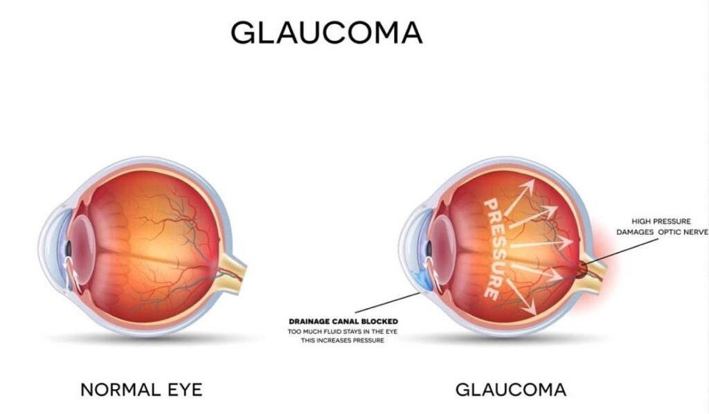
To understand glaucoma, you must understand fluid dynamics. Your eye constantly produces a fluid called aqueous humor, which provides nutrients to the eye’s structures. This fluid must drain at the exact same rate it is produced through a drainage angle structure termed the trabecular meshwork.
If this drain becomes blocked or sluggish, fluid builds up. This elevates your Intraocular Pressure (IOP). Prolonged high pressure physically compresses and destroys the delicate fibers of the optic nerve at the back of the eye. Raised IOP also restricts blood supply to the nerve head resulting in tissue damage.
The Primary Types of Glaucoma in Singapore
- Primary Open-Angle Glaucoma: The most common form globally. The drainage angle remains “open,” but the meshwork itself becomes inefficient over time. It is entirely painless and symptomless in early stages.
- Angle-Closure Glaucoma: Highly prevalent among East Asians due to our specific ocular anatomy (shallower anterior chambers). The iris can physically oppose the drainage angle, gradually or acutely blocking the drainage angle. Acute angle-closure causes a rapid, severe pressure spike and is a medical emergency.
- Normal-Tension Glaucoma: A variant where optic nerve damage occurs despite the eye pressure remaining within the “normal” range (under 21 mmHg). It is often linked to poor blood flow to the optic nerve.
Treatment Options: Medical, Laser, and Surgery
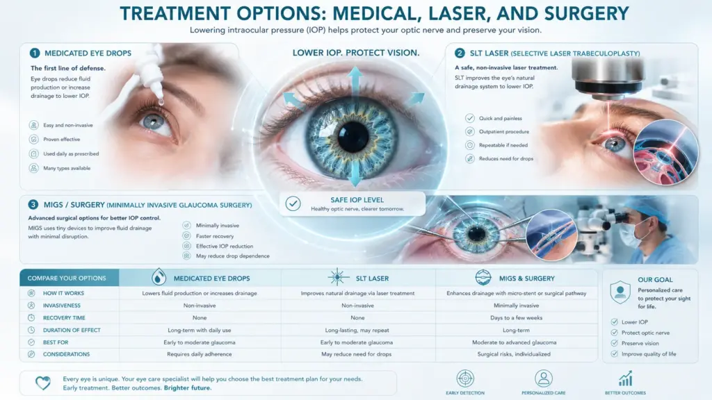
While vision lost to glaucoma cannot be restored, we can halt or significantly slow its progression by lowering the IOP to a safe target range.
| Management Option | How It Works | Clinical Profile & Considerations | Singapore MediSave / Cost Context |
| Medicated Eye Drops | Decreases aqueous production or increases drainage. | Pros: Non-invasive outpatient management. Cons: Requires strict daily compliance; can cause ocular surface redness or dry eye. | Out-of-pocket expense; not claimable under MediSave for outpatient medication. |
| SLT Laser (Selective Laser Trabeculoplasty) | Applies gentle laser energy to the drainage tissue to stimulate better fluid outflow. | Pros: Evidence-based first-line alternative to daily drops; very safe profile. Cons: Effect may wear off after a few years (though it can be repeated). | MediSave Claimable under specific surgical table limits. |
| MIGS & Trabeculectomy (Surgery) | Creates a new drainage pathway or inserts microscopic stents/tube to bypass the blockage. | Pros: Effective for moderate to severe cases or when drops fail. Cons: Carries standard intraocular surgical risks (infection, bleeding). | MediSave Claimable. Integrated Shield Plans may cover depending on policy. |
If you experience severe, throbbing eye pain, a one-sided headache, nausea, blurred vision, and rainbow-colored halos around lights, do not wait. This indicates an acute angle-closure attack. Immediate medical intervention is required to prevent rapid, permanent blindness. Proceed to your nearest A&E or contact our clinic immediately.
Screening Guidelines: Your Next Steps
Age and genetics are significant risk factors. If you have a direct family member with glaucoma, your risk increases up to ninefold.
The American Academy of Ophthalmology (AAO) recommends a baseline comprehensive eye evaluation at age 40, even if you have no symptoms. For individuals over 65, or those with high myopia, diabetes, or a family history, screening should occur every 1 to 2 years.
Do not wait for your vision to fade. Protect your optic nerve through early, precise diagnostic screening.